16. Skeleton and Movements - part 05 - Appendicular skeleton and Disorders related to muscles
16. Skeleton and Movements - part 05 - Appendicular skeleton and Disorders related to muscles
B. Appendicular skeleton :
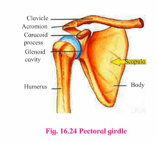
Pectoral girdle :
Clavicle :
Scapula :
Bones of forelimb :
Humerus :

Radius and Ulna :

Carpals :
Metacarpals :

Phalanges :
Pelvic girdle :

Bones of lower limb :
Femur :

Tibia and fibula :

Tibia :

Fibula :
Tarsals :
Metatarsals :

Phalanges :
Types of joints :
Classification of Fibrous joints
Sutures :


2. Cartilagenous or slightly movable joints :
a. Synchondroses :

b. Symphysis :
3. Synovial joints or freely movable joints :
Pivot joint :

Ball and socket joint :

Hinge joint :
Condyloid joint :

Gliding joint :
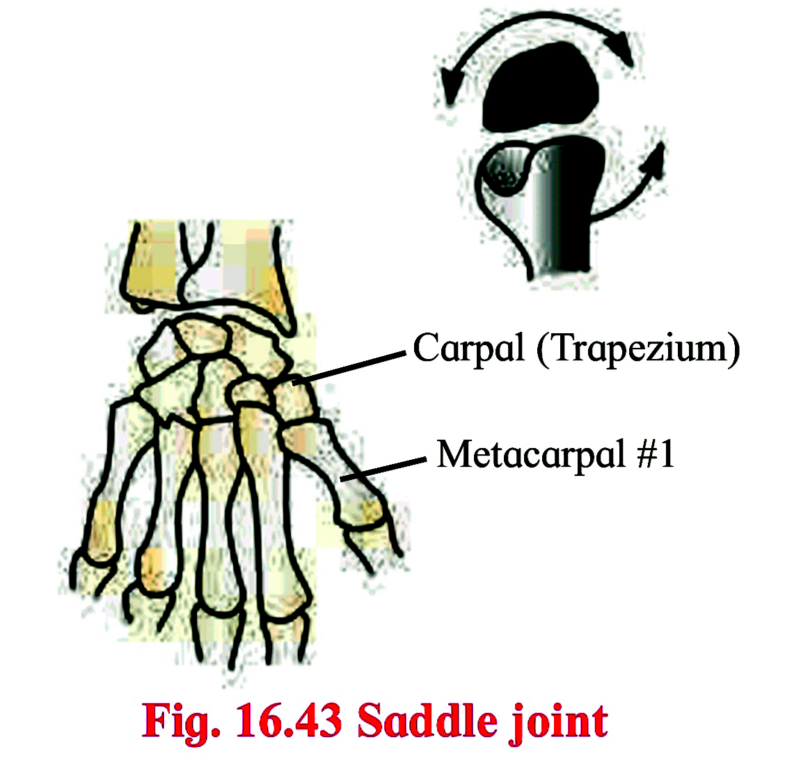
Saddle joint :
Disorders related to muscles :
Muscular dystrophy :
Myasthenia gravis :
Disorders related to bones :
Arthritis :
i. Osteoarthritis :
ii. Gouty arthritis (Gout) :
iii. Rheumotoid arthritis :
Osteoporosis :
- Appendicular skeleton consists of bones of limbs and girdles.
Pectoral girdle :

- Shoulder girdle
- It attaches forelimb skeleton with axial skeleton.
- There are two pectoral girdles, each consists of a shoulder blade or scapula and collar bone or clavicle.
Clavicle :
- It is ‘s’ shaped slender bone.
- One end of clavicle is attached to acromion process of scapula.
- The other rounded end called sternal end attaches to manubrium of sternum. This connects upper arm skeleton to axial skeleton.
Scapula :
- It is a large, flat, triangular bone that occupies posterior chest wall extending from second to seventh ribs.
- It is attached to axial skeleton by muscles and tendons.
- At it’s lateral angle, scapula bears a concave socket called glenoid cavity.
- Head of humerus (the upper arm bone) fits into the glenoid cavity.
- Two processes arise from scapula, a beak like coracoid process that projects from lateral angle of scapula and acromion process, easily felt as high point of shoulder.
- Both are meant for attachment of muscles.
Bones of forelimb :
- It consists of humerus, radius and ulna (together forming forearm bones), Bones of wrist -the carpals, bones of palm-the metacarpals and bones of digitsphalanges
- Together making to 30 bones.
Humerus :

- This is the bone of upper arm. It has hemispherical head at it's proximal end.
- On either side of head of humerus are present a pair of projections termed greater and lesser tubercles.
- There is a deep groove between the tubercles called bicipital groove where a tendon of biceps muscle is attached.
- Shaft of humerus shows deltoid tuberosity.
- Distal end of humerus shows pulley like part called trochlea that articulates with ulna.
Radius and Ulna :

- Radius is located laterally on thumb side of the forearm.
- Proximal end of radius has disc like head that articulates with humerus bone.
- The shaft of radius widens distally to form styloid process.
- Ulna is located medially on little finger side of forearm.
- At the proximal end of ulna there is a prominent process called 'Olecranon process’ that forms elbow joint with humerus bone.
- On the lateral side, near the upper end of ulna is present the radial notch into which the side of head of radius is fixed.
- Radius and ulna articulate with each other at upper and lower extremities by superior and inferior radio-ulnar joints.
- In between the shaft of two bones, interosseous membrane is present.
Carpals :
- These are bones of wrist, arranged in two rows of four each.
Metacarpals :

- Five elongated metacarpals form bones of palm.
- Their proximal ends join with carpals and distal ends form knuckles.
Phalanges :
- These are bones of fingers and thumb.
- Four fingers have three phalanges each and thumb has two; thus making it fourteen phalanges in each hand.
Pelvic girdle :

- Pelvic or hip girdle connects hind limb skeleton with axial skeleton.
- It is made up of two hip bones called coxal bones.
- They unite posteriorly with sacrum.
- Each large irregularly shaped bone, the coxal bone is made up of three parts, ilium, ischium and pubis.
- At the point of fusion of three bones, a cavity called acetabulum is present that forms ball and socket joint with thigh bone.
- The two pubis bones are joined medially by cartilaginous joint called pubic symphysis.
- Pubis and ischium together form a ring of bone that encloses a space called obturator foramen.
Bones of lower limb :
Femur :

- The thigh bone is the longest a bone in the body.
- The head is joined to shaft at an angle by a short neck.
- It forms ball and socket joint with acetabulum cavity of coxal bone.
- The lower one third region of shaft is triangular flattened area called popliteal surface.
- Distal end has two condyles that articulate with tibia and fibula.
Tibia and fibula :

- These are the two long bones of shank or lower leg.
- The two are connected to each other at the extremities.
- In between the two bones interosseous membrane is present.
Tibia :

- It is much thicker and stronger than fibula.
- It’s broad and expanded upper end articulates with femur.
- Lower end articulates with talus, a tarsal bone.
Fibula :
- It is a long slender bone on lateral side of tibia
Tarsals :
- These are the bones of ankle.
- Seven tarsals are arranged in three row, two proximal,one intermediate and four distal.
Metatarsals :

- Five metatarsal bones support the sole region of the foot.
- Proximally they attach with distal row of tarsals.
- Distally metatarsals articulate with phalanges.
Phalanges :
- These are the bones of the toes.
- Except the big toe which has two phalanges, rest four toes have three phalanges each.
Types of joints :
- A point where two or more bones get articulated is called joint or articulation or arthrosis.
- Study of joints is called arthrology.
- Though bones are rigid, the ligaments that cover the bones, forming a joint render slight flexibility to the bones.
- fibrous joints/ synarthroses / immovable joints
- cartilagenous / slightly movable joints/amphiarthroses
- synovial / freely movable / diarthroses
- fibrous joints/ synarthroses / immovable joints
- In this joint, the articulating bones are held together by means of fibrous connective tissue.
- Bones do not exhibit movement. Hence it is immovable or fixed type of joint.
Classification of Fibrous joints
- Sutures,
- Syndesmoses
- Gomphoses.
Sutures :


- It is composed of thin layer of a dense fibrous connective tissue.
- Sutures are places of growth.
- They remain open till growth is complete.
- On completion of growth they tend to ossify.
- Sutures may permit some moulding during childhood.
Classification of sutures :
Syndesmoses :
- It is present where there is greater distance between articulating bones.

- At such locations, fibrous connective tissue is arranged as a sheet or bundle.
- e.g. Distal tibiofibular ligament, inter osseous membrane between tibia and fibula and that between radius and ulna.

Gomphoses :

- In this type of joint a cone shaped bone fits into a socket provided by other bone.
- e.g. Tooth and jow bones.
2. Cartilagenous or slightly movable joints :
- These are also called as amphiarthroses
- These joints are neither fixed nor freely movable.
- Articulating bones are held together by hyaline or fibrocartilages.
- They are further classified as
a. Synchondroses
b. Symphysis
a. Synchondroses :

- The two bones are held together by hyaline cartilage.
- They are meant for growth.
- On completion of growth, the joint gets ossified.
- Example: Epiphyseal plate found between epiphysis and diaphysis of a long bone, Rib – Sternum junction.
b. Symphysis :
- In this type of joint, broad flat disc of fibrocartilage connects two bones.
- These occur in midline of the body.
- e.g. intervertebral discs.
3. Synovial joints or freely movable joints :
- They are also called as diarthroses.
- It is characterized by presence of a space called synovial cavity between articulating bones that renders free movement at the joint.

- Articulating surfaces of bones at a synovial joint are covered by a layer of hyaline cartilage.
- It reduces friction during movement and helps to absorb shock.
- Synovial cavity is lined by synovial membrane that forms synovial capsule.
- Synovial membrane secretes synovial fluid.
- Synovial fluid is a clear, viscous, straw coloured fluid similar to lymph.
- It is viscous due to hyaluronic acid.
- Fluid also contains nutrients, mucous and phagocytic cells to remove microbes.
- Synovial fluid lubricates the joint, absorbs shocks, nourishes the hyaline cartilage and removes waste materials from hyaline cartilage cells (as cartilage is avascular) phagocytic cell destroy microbes and cellular debris formed by wear and tear of the joint.
- If the joint is immobile for a while, the synovial fluid becomes viscous and as joint movement starts, it becomes less viscous.
- The joint is provided with capsular ligament and numerous accessory ligaments.
- The fibrous capsule is attached to periosteum of articulating bones.
- The ligament helps in avoiding dislocation of joint.
- Let us study types of synovial joints.
- Note that any type of synovial joint will show above mentioned components.

Pivot joint :

- Here, the rounded or pointed surface of one bone articulates with a ring formed partly by another bone and partly by ligament.
- Rotation only around it’s own longitudinal axis is possible.
- Example : In joint between atlas and axis vertebrae, head turns side ways to form ‘NO’ joint.
Ball and socket joint :

- Ball like surface of one bone fits into cup like depression of another bone forming a moveble joint.
- Multiaxial movements are possible.
- This type of joint allows movements along all three axes and in all directions.
- Example : Shoulder and hip joint
Hinge joint :
- In a hinge joint, convex surface of one bone fits into concave surface of another bone.

- In most hinge joints one bone remains stationery and other moves.
- Angular, opening and closing motion like that of a hinge is possible.
- In this joint only monoaxial movement takes place like flexion and extension.
- Example: Elbow and knee joint.

Condyloid joint :

- It is an ellipsoid joint.
- The convex oval shaped projection of one bone fits into oval shaped depression in another bone.
- It is a biaxial joint because it permits movement along two axes viz. flexion, extension, abduction, adduction and circumduction is possible.
- Example : Metacarpophalyngeal joint
Gliding joint :
- A planar joint, where articulating surfaces of bones are flat or slightly curved.
- These joints are non-axial because motion they allow does not occur along an axis or a plane.
- Example : Intercarpal and intertarsal joints.
Saddle joint :
- This joint is a characteristic of Homo sapiens.

- Here, articular surface of one bone is saddle-shaped and that of other bone fits into such saddle as a sitting rider would sit.
- Each bone has both concave and convex areas.
- It is a modified condyloid joint in which movement is somewhat more free.
- It is a biaxial joint that allows flexion, extension, abduction, adduction and circumduction.
- Example : Carpometacarpellar jont between trapezium carpal and metacarpal of thumb.
Disorders related to muscles :
Muscular dystrophy :
- It is a gradual wasting disease affecting various groups of muscles.
- Genetically inherited in families.
- Usually voluntary skeletal muscles are weakened whereas internal muscles such as diaphragm are not affected.
- Duchenne type usually occurs in boys affecting lower limbs.
- Limb girdle muscular dystrophy affects the muscles of shoulders or hips and it usually starts in adults of 20-35 years.
- No treatment appears to cure the disease.
Myasthenia gravis :
- It is a weakness of skeletal muscles.
- It is caused by an abnormality at the neuromuscular junction that partially blocks contraction.
- It is an autoimmune disorder caused by an excess of certain antibodies in the blood stream.
- Antibodies bind to accetylecholine receptors of neuromuscular junction.
- Thus transmission of nerve impulses to the muscle fibres is blocked.
- This causes progressive and extensive muscle weakness.
- It may affect the eye and eyelid movements, facial expression and swallowing.
- The degree of muscle weakness varies form local to general.
- Example of symptoms are – Ptosis, (diplopia or double vision) difficulty in swallowing,chewing and speech.
Disorders related to bones :
Arthritis :
- It is an inflammation of joints.
- It is a painful disorder of bones, ligaments tendons etc.
- In this disorder, joints become swollen, stiff and painful.
- It can lead to disability. Arthritis is of three types.
i. Osteoarthritis :
- In this, joint cartilage is degenerated.
- It is caused by various factors like aging, obesity, muscle weakness, etc.
- This is most common type of arthritis that affects hands, knees and spine.
ii. Gouty arthritis (Gout) :
- In this disorder joint pain occurs due to deposition of uric acid in joints.
- If uric acid is produced in excess or is not excreted, it accumulates in joints as sodium urate and degenerates cartilage, causing inflammation and pain.
- It generally affects joints of feet.
iii. Rheumotoid arthritis :
- It is an autoimmune disorder where body’s immune system attacks it’s own tissues.
- In rheumatoid arthritis, synovial membrane swells up and starts secreting extra synovial fluid. This fluid exerts pressure on joint and makes it painful.
- Membrane may develop abnormal granulation tissue called pannus.
- Pannus may erode cartilage.
- Fibrous tissue gets ossified and may lead to stiffness in joints.
Osteoporosis :
- In this disorder, bones become porous and hence brittle.
- It is primarily age related disease more common in women than men.
- As age advances, bone resorption outpaces bone formation hence bones loose mass and become brittle. More calcium is lost in urine, sweat etc. than it is gained through diet.
- Hence prevention of disease is better than treatment by consuming adequate amount of calcium and exercise at young age.
- Osteoporosis may be caused due to decreasing estrogen secretion after menopause, deficiency of vitamin D,low calcium diet, decreased secretion of sex hormones and thyrocalcitonin.
- Apart from fractures, osteoporosis may lead to shrinkage of vertebrae, height loss, hunched back and bone pain.


Comments
Post a Comment